Fluoroscopy is a form of real-time X-RAY that employs a fluoroscope to examine the tissues and deep structures within the body. A fluoroscope is a device that projects radiographic images in a movie-like sequence onto a monitor. It is often used with contrast material which clearly defines the area being examined by making it appear darker. Fluoroscopy makes it possible to see internal organs in motion.
Fluoroscopy Procedures Offered:
- Hysterosalpingography (HSG)
HSG
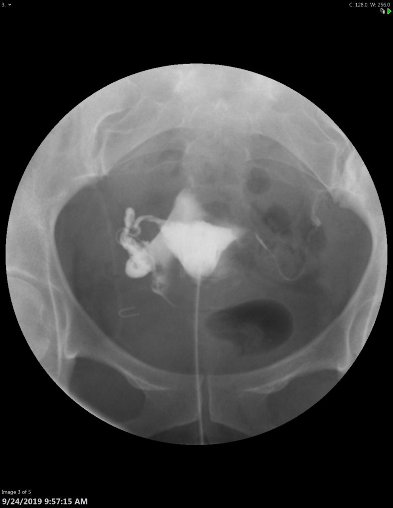
HSG uses Fluoroscopy to examine the uterus and fallopian tubes of a woman who is having difficulty becoming pregnant. It’s also used to investigate miscarriages resulting from abnormalities within the uterus and to determine the presence and severity of adhesions or uterine fibroids tumor masses, adhesions, and uterine fibroids. Contrast material is administered through a catheter.
During the Exam
During the exam, you will be positioned on the exam table, with your knees bent or your feet held up with stirrups. A speculum is inserted into the vagina, similar to a routine gynecological exam. The cervix is then cleansed, and a catheter is inserted into the cervix. The speculum is removed and you will be carefully positioned underneath the Fluoroscopy camera. The contrast material then begins to fill the uterine cavity, fallopian tubes, and peritoneal cavity through the catheter and fluoroscopic images are taken and stored for further review.
Preparation
You may be asked to undress whatever part of your body needs examination. You may also be asked to remove jewelry, eyeglasses and any metal objects.
The hysterosalpingography procedure is best performed one week after menstruation but before ovulation to make certain that you are not pregnant during the exam. You should inform your physician of any medications being taken and if there are any allergies, especially to iodinated contrast materials.