Mammography is a specific type of breast imaging that uses low-dose Digital Radiography (X-RAY) to detect cancer early – before a woman experiences symptoms – when it is the most treatable or evaluate abnormal clinical findings – such as a breast lump or nipple discharge – that have been found by a woman and her doctor.
UDMI is pleased to announce that we have partnered with ScreenPoint Medical to offer Transpara® Artificial Intelligence (AI) as an add-on to your annual screening mammogram.**
Finding breast cancer early makes it easier to treat. That’s why it’s really important for all women to have access to the newest tools that can find the disease early. Around the world, about 1 in 8 women will get breast cancer in their lifetime, but with regular check-ups and early detection, many can survive it.
Transpara® AI is a smart tool that uses advanced AI technology to help your doctors when they look at your mammogram. Studies show that when your doctors use Transpara® AI, they can find more cancers than if they check alone. Transpara® AI is not meant to take the doctors’ place but to help them find cancers sooner. No additional radiation or exam time is necessary. AI is used by the physicians while reviewing the images following the exam.
**There is a $20 fee (payable on date of service) to use Artificial Intelligence for your screening (at this time, the fee is not covered by insurance). UDMI can still do your mammogram if you choose to not use AI.
Mammography can be used for either screening or diagnostic purposes:
- Screening Mammography – an annual exam used to detect breast changes in women who have no signs or symptoms or readily apparent breast abnormalities.
- Diagnostic Mammography – used to investigate suspicious breast changes, such as a new lump, breast pain, an unusual skin appearance, nipple thickening or nipple discharge. It’s also used to evaluate abnormal findings on a screening mammogram.
UDMI is fully equipped with the latest technology in Mammography known as 3-dimensional (3D) digital breast tomosynthesis, or 3D Mammography.
3D tomosynthesis is different from a standard mammogram. Standard Mammography includes two Digital Radiography (X-RAY) of each breast from different angles. Standard mammograms are very effective, but are limited by dense breasts and overlapping tissue. 3D tomosynthesis overcomes these issues by taking multiple images of each breast allowing clearer visualization of the breast tissue through multiple layers and angles collectively resulting in a 3D image.
In 2019, the American Society of Breast Surgeons (ASBrS) issued new breast screening guidelines which include a recommendation that all screenings be done with 3D tomosynthesis. If you choose, however, to not undergo a tomosynthesis exam, a state-of-the-art standard digital 2D mammogram will still be performed.
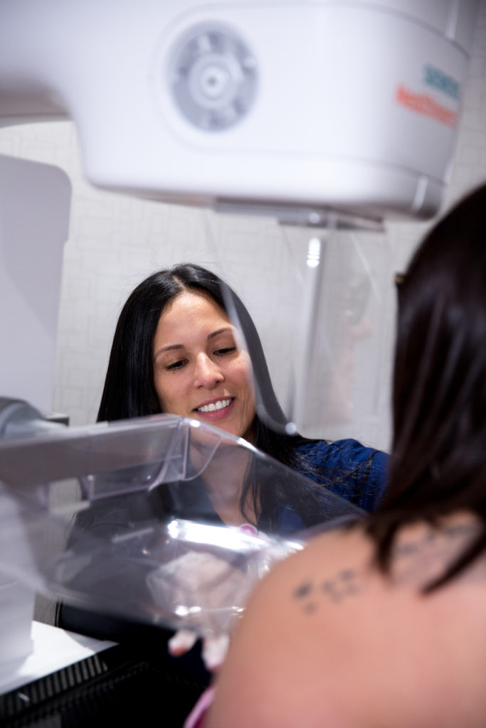
During the Exam
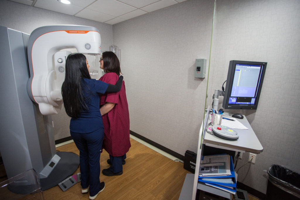
During a mammogram, your breasts are compressed between two firm surfaces to spread out the breast tissue. UDMI’s new machine is designed to be more comfortable with less compression than the traditional 2D mammogram but you may still feel slight discomfort or pressure. An X-RAY captures black-and-white images of your breasts that are displayed on a computer screen and examined by a doctor who looks for signs of cancer. During the X-RAY exposure you will be asked to hold your breath and stand still.
After the images are taken you may be asked to wait while our on-site radiologist reviews the images. The entire procedure usually takes less than 15 minutes.
Preparation
Screening exams can be scheduled for a time when your breasts are less likely to be tender (usually the week before or the week after your menstrual period). However, if a new breast lump is identified either by self or clinical breast exam, the exam should be scheduled as soon as possible regardless of tenderness.
If your prior mammogram was performed outside UDMI, be sure to bring your prior images with you for comparison (you can request the prior facility to put the images on a disc). The radiologist reviewing your mammogram will not be able to detect changes from the prior mammogram without the prior images.
Dress in a two-piece outfit as you will be asked to remove your shirt and bra and change into a gown. Don’t use deodorants, antiperspirants, powders, lotions, creams or perfumes under your arms or on your breasts. Many women find it helpful to take over-the-counter pain medication about an hour before their mammogram to ease the discomfort of the test.